
The pulse is the palpable throbbing sensation you feel over the peripheral arteries. It occurs as a result of rapid blood flow within the arteries during the contraction of the heart.
Quiz Your Musculoskeletal System Knowledge Here
There are 9 common pulse points on the body. You can feel them by lightly palpating the artery against the underlying bone or muscle.
Monitoring pulse is a crucial part of physical assessment and observing vital signs. The characteristics (eg: abnormally slow, fast, or irregular pulse) of the pulse will give you an indication of the cardiovascular system’s ability to deliver enough blood to the body.
The radial pulse you feel from the radial artery is the most common pulse point for taking pulse in adolescents and adults.
But during a cardiovascular collapse, radial pulse usually will not be palpable. In this case, a more central site like the carotid artery should be used to check for pulse.
This article covers:
- definition of pulse,
- characteristics of pulse,
- normal pulse rate,
- factors influencing pulse rate,
- 9 common pulse points on the body, and
- how to locate pulse sites
Definition of the pulse
The pulse can be defined as a rhythmic wave of pressure formed by the expansion of an artery due to the heart’s contraction of the left ventricle.

Human Anatomy & Physiology Course
- Guidebooks
- Complete Diagrams & Lessons
- Certifications
- Quiz & Answers
- And More…
Characteristics of the pulse
When you assess the pulse, you are observing for characteristics of the pulse. The pulse characteristics offers valuable data for determining the integrity of the client’s cardiovascular system.
Key characteristics of the pulse include:
- Frequency or pulse rate
- Rhythm or regularity
- Strength (amplitude) or intensity
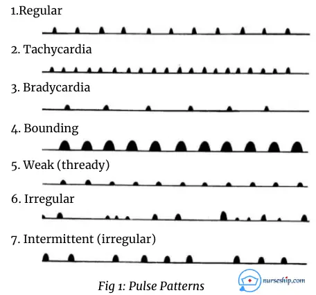
1. Frequency or Pulse Rate
The pulse rate is the pulsation you feel for one minute. The pulse rate should be within the normal range.
See Table 2 for normal pulse rate as per age.
when the pulse rate is below the normal range (less than 60 in adults) is called Bradycardia.
When the pulse rate is above the normal range (more than 100 in adults) is called Tachycardia.
Pulse is documented as pulse beats per minute (bpm). To determine pulse rate, you have to count pulse for 60 seconds.
For instance, you can write pulse rate as 74/min or 74 bpm.
Note: Always refer your institute’s guideline when documenting.
2. Rhythm or Regularity of Pulse
Rhythm is the time interval between pulse beats.
You assess the rhythm of the pulse to determine whether it’s regular or irregular.
Normal pulse beats at regular intervals.
Irregular pulse indicates irregular contractions of the heart. Then pulse must be assessed bilaterally. Also you refer the client to a senior clinician for further evaluation.
3. Strength (amplitude) or Intensity of Pulse
The strength of the pulse is the force of blood felt at each heartbeat.
It is determined by the amount of blood pushed out of the heart into the arteries with every heartbeat.
The force will be at normal strength if the client has a normal pulse.
Abnormal strengths of the pulse are:
- A weak or feeble pulse indicates reduced cardiac output and requires immediate action.
- A strong, bounding pulse indicates high blood pressure.
The strength of the pulse also should be graded on a scale of 0 to 4 +.
| Grade | Indication |
| 0 | No pulse |
| 1+ | weak, feeble, faint but detectable pulse |
| 2+ | slightly more diminished pulse than normal |
| 3+ | normal pulse |
| 4+ | bounding pulse |
Factors that Affect Pulse Rate
- Age
- Gender
- Physique
- Body temperature
- Hemorrhage
- Exercise
- Emotional status
- Blood pressure
- Pain
- Food intake
- Medications (stimulants increase the pulse rate and depressants decrease the pulse rate)
Normal Pulse Rate by Age
| Age | Normal pulse rate (bpm) |
| Less than 1 month | 120 – 160 |
| 1 to 12 months | 80 – 140 |
| 12 to 24 months | 80 – 130 |
| 2 – 6 years | 75 – 120 |
| 6 – 12 years | 75 – 110 |
| 12 years above | 60 – 100 |
Related Article: Hemodynamic Monitoring
Challenge Yourself With Our Quizzes
9 Common Pulse Points
9 most commonly assessed pulse points on the body by nurses are:
- Temporal pulse – over the temple
- Carotid pulse – at the side of the neck
- Apical pulse – over the 5th intercostal space (ICS) at left mid-clavicular line.
- Brachial pulse – on the antecubital fossa (crook) of the arm
- Radial pulse – in the wrist below the thumb
- Femoral pulse – in the groin
- Popliteal pulse – behind the knee
- Posterior tibial pulse – to the side of the ankle
- Dorsalis pedis pulse – on the front of the foot.
1. Temporal artery pulse
The superficial temporal artery is where you assess temporal pulse with your index and middle fingertips.
It can be located over the temple just in front of the tragus of the external ear.
Why is temporal pulse measured?
The temporal artery pulse site is assessed during nursing head-to-toe assessment
Also, this pulse site is easily accessible in children.
2. Carotid artery pulse
The anatomical location of the carotid pulse is along the medial edge of the sternocleidomastoid muscle in the neck (i.e., mid-line between earlobe and chin below the jawline.)
Use index and middle fingertips to palpate carotid artery.
When you assess carotid pulse, be cautious and feel both sides separately to avoid vagus nerve stimulation. Because it will reduce the heart rate and circulation to the brain.
Why is the carotid pulse measured?
The carotid artery pulse site is used:
- to assess peripheral pulse characteristics
- to assess the presence of pulse when a person collapses and during cardiopulmonary resuscitation (CPR)
- when other peripheral pulses are not palpable.
3. Apical pulse
The apical pulse can be anatomically located over the 5th intercostal space at the left mid-clavicular line.
Unlike other pulses, the apical pulse is unilateral and auscultated directly over the apex of the heart.
Always count apical pulse for 1 full minute.
Why is the apical pulse measured?
The apical pulse site is assessed during the head-to-toe assessment, instead of palpating you listen to the apical pulse using the stethoscope.
Apical pulse is the preferred site to measure heart rate in newborns and infants.
4. Brachial artery pulse
The brachial pulse is felt on the anterior aspect of the elbow by gently pressing the artery against the underlying bone with the middle and index fingers (i.e: the groove between the biceps and triceps at the antecubital fossa).
Why is the brachial pulse measured?
The brachial artery pulse site is used:
- to measure blood pressure with a stethoscope and sphygmomanometer.
- to assess the circulation of the lower arm
5. Radial artery pulse
The anatomical position of the radial pulse is immediately above the wrist joint near the base of the thumb.
This is the most commonly measured pulse by nurses to assess peripheral pulse characteristics.
The radial artery pulse can be felt by gently pressing the radial artery against the underlying bone with the middle and index fingers.
Why is radial pulse measured?
Radial artery pulse site is assessed:
- to check blood circulation of the hand
- to check peripheral pulse characteristics
Click here for a demonstration of how to take radial pulse.
6. Femoral artery pulse
The femoral pulse is anatomically located below the inguinal ligament between the pubic symphysis and anterior superior iliac spine.
Femoral artery pulse are another major pulse you may palpate during shock or cardiac arrest.
Why is femoral pulse measured?
The femoral artery pulse site is assessed:
- to check blood circulation of the leg
- to check pulse during a shock and cardiac arrest
7. Popliteal artery pulse
The popliteal pulse is located behind the knee in popliteal fossa.
This pulse is more difficult to palpate as compared to other pulse sites.
Why is popliteal pulse measured?
Popliteal artery pulse site is assessed:
- to auscultate lower limbs blood pressure
- to check blood circulation of the lower leg
8. Posterior tibial artery pulse
The posterior tibial pulse is anatomically located inner side of the ankles below the medial malleolus.
You can palpate the tibial artery by gently pressing against the underlying bone with the middle and index fingers.
Why is posterior tibial pulse measured?
The posterior tibial artery pulse site is assessed to check blood circulation of the foot
9. Dorsalis pedis pulse
The anatomical location of the dorsalis pedis pulse is the groove between the first and second toes slightly medial on the top of the foot.
Why is dorsalis pedis pulse measured?
Dorsalis pedis artery pulse site is also assessed to check blood circulation of the foot
Take Our Nursing Fundamentals Quiz Here – Assess Your Knowledge
Conclusion
Pulse is an important general vital sign. The number of pulsing sensations you feel during 1 minute is the pulse rate per minute. And several factors may affect the pulse rate.
The characteristics of the pulse give information about the status of the cardiovascular system.
There are 9 common pulse points on the surface of the body. Namely, temporal pulse, carotid pulse, apical pulse, brachial pulse, radial pulse, femoral pulse, popliteal pulse, posterior tibial pulse, and dorsalis pedis pulse.
Click here to learn how to take pulse.
Reference
Hilton, P. (2005). Fundamental nursing skills. London: Whurr Publishers.
Perry, A.G., Potter, P.A., Ostendorf, W.R. (2018). Clinical nursing skills & techniques (9th ed.). Elsevier: St. Louis.
Lynn, P., & Taylor, C. (2011). Taylor’s clinical nursing skills (3rd ed.). Philadelphia: Wolters Kluwer Health/Lippincott Williams & Wilkins.