In this article we’ll learn about 12 cranial nerves and their anatomy, function, and how to assess them.
The cranial nerves are a set of twelve nerves that emerge directly from the brain and pass through the openings in the skull to reach various parts of the head, neck, and trunk.
They are responsible for the sensory and motor functions of the head and neck, and they play a crucial role in many of the body’s physiological processes.
Overview of the 12 cranial nerves
Here is a brief overview of the 12 cranial nerves.
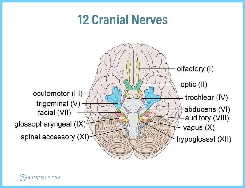
Olfactory nerve (I)
This nerve is responsible for the sense of smell.
It originates in the olfactory epithelium of the nasal cavity and passes through the cribriform plate of the ethmoid bone to reach the olfactory bulb.
Optic nerve (II)
This nerve is responsible for vision.
It originates in the retina of the eye and passes through the optic canal of the sphenoid bone to reach the brain.
Oculomotor nerve (III)
This nerve controls the movement of most of the eye muscles, including the superior, inferior, and medial rectus muscles, and the inferior oblique muscle. It also controls the size of the pupil and the shape of the lens of the eye.
It originates in the midbrain and passes through the superior orbital fissure.
Trochlear nerve (IV)
This nerve controls the movement of the superior oblique muscle of the eye.
It originates in the midbrain and passes through the superior orbital fissure.
Trigeminal nerve (V)
This nerve has three branches that provide sensory and motor innervation to the face, scalp, and teeth. It also controls the muscles of mastication.
It originates in the pons and passes through the superior orbital fissure, foramen rotundum, and foramen ovale.
Abducens nerve (VI)
This nerve controls the movement of the lateral rectus muscle of the eye.
It originates in the pons and passes through the superior orbital fissure.
Facial nerve (VII)
This nerve controls the muscles of facial expression, taste sensation in the anterior two-thirds of the tongue, and lacrimal and salivary glands. It also provides sensory innervation to the external ear.
It originates in the pons and passes through the internal acoustic meatus, facial canal, and stylomastoid foramen.
Vestibulocochlear nerve (VIII)
This nerve is responsible for hearing and balance.
It has two branches: the vestibular branch, which provides sensory information about balance and spatial orientation, and the cochlear branch, which provides sensory information about hearing.
It originates in the inner ear and passes through the internal acoustic meatus.
Glossopharyngeal nerve (IX)
This nerve controls the muscles of the pharynx and provides taste sensation in the posterior one-third of the tongue. It also provides sensory innervation to the carotid body and carotid sinus.
It originates in the medulla oblongata and passes through the jugular foramen.
Vagus nerve (X)
This nerve provides parasympathetic innervation to the thoracic and abdominal organs, controls the muscles of the larynx and pharynx, and provides sensory innervation to the external ear and some viscera.
It originates in the medulla oblongata and passes through the jugular foramen.
Accessory nerve (XI)
This nerve controls the sternocleidomastoid and trapezius muscles.
It originates in the spinal cord and passes through the jugular foramen.
Hypoglossal nerve (XII)
This nerve controls the muscles of the tongue.
It originates in the medulla oblongata
Here is a table summarizing the information on the 12 cranial nerves.
| Cranial Nerve | Function | Anatomy | Type |
| I – Olfactory nerve | Sense of smell | Originates in the olfactory epithelium of the nasal cavity and passes through the cribriform plate of the ethmoid bone to reach the olfactory bulb | Sensory |
| II – Optic nerve | Vision | Originates in the retina of the eye and passes through the optic canal of the sphenoid bone to reach the brain | Sensory |
| III – Oculomotor nerve | Eye movement, pupil constriction, lens shape | Originates in the midbrain and passes through the superior orbital fissure | Motor |
| IV – Trochlear nerve | Eye movement | Originates in the midbrain and passes through the superior orbital fissure | Motor |
| V – Trigeminal nerve | Sensation to the face, scalp, and teeth, controls muscles of mastication | Originates in the pons and passes through the superior orbital fissure, foramen rotundum, and foramen ovale | Both sensory and motor |
| VI – Abducens nerve | Eye movement | Originates in the pons and passes through the superior orbital fissure | Motor |
| VII – Facial nerve | Muscles of facial expression, taste sensation in anterior two-thirds of the tongue, lacrimal and salivary glands, sensory innervation to external ear | Originates in the pons and passes through the internal acoustic meatus, facial canal, and stylomastoid foramen | Both sensory and motor |
| VIII – Vestibulocochlear nerve | Hearing and balance | Originates in the inner ear and passes through the internal acoustic meatus | Sensory |
| IX – Glossopharyngeal nerve | Muscles of the pharynx, taste sensation in posterior one-third of the tongue, sensory innervation to carotid body and carotid sinus | Originates in the medulla oblongata and passes through the jugular foramen | Both sensory and motor |
| X – Vagus nerve | Parasympathetic innervation to thoracic and abdominal organs, muscles of larynx and pharynx, sensory innervation to external ear and some viscera | Originates in the medulla oblongata and passes through the jugular foramen | Both sensory and motor |
| XI – Accessory nerve | Sternocleidomastoid and trapezius muscles | Originates in the spinal cord and passes through the jugular foramen | Motor |
| XII – Hypoglossal nerve | Muscles of the tongue | Originates in the medulla oblongata | Motor |
Mnemonic to remember 12 cranial nerves and their function
There are several mnemonics that can be used to remember the 12 cranial nerves. Here is one of those mnemonics to easily remember 12 cranial nerves in order.
“On Old Olympus Towering Tops, A Finn And German Viewed Some Hops“
Each word in this sentence represents the first letter of cranial nerves in order along with their roman numeral.
| Cranial Nerve | Mnemonic |
| I – Olfactory nerve | On |
| II – Optic nerve | Old |
| III – Oculomotor nerve | Olympus |
| IV – Trochlear nerve | Towering |
| V – Trigeminal nerve | Tops |
| VI – Abducens nerve | A |
| VII – Facial nerve | Finn |
| VIII – Vestibulocochlear nerve | And |
| IX – Glossopharyngeal nerve | German |
| X – Vagus nerve | Viewed |
| XI – Accessory nerve | Some |
| XII – Hypoglossal nerve | Hops |
Additionally, to remember the functions of each nerve, another mnemonic can be added.
“On Old Olympus Towering Tops, A Finn And German Viewed Some Hops. Very Good, Such Heaven!”
The last sentence “Very Good, Such Heaven!” is an additional phrase that can be used to remember the functions of each cranial nerve.
- Cranial nerves I-IV: sensory nerves involved in vision, hearing, smell, and eye movement.
- Cranial nerves V-XII: mixed nerves involved in sensory, motor, and autonomic functions of the head, neck, and viscera.
| Cranial Nerve | Mnemonic | Function |
| I – Olfactory nerve | On | Sense of smell |
| II – Optic nerve | Old | Vision |
| III – Oculomotor nerve | Olympus | Eye movement, pupil constriction, lens shape |
| IV – Trochlear nerve | Towering | Eye movement |
| V – Trigeminal nerve | Tops | Sensation to the face, scalp, and teeth, controls muscles of mastication |
| VI – Abducens nerve | A | Eye movement |
| VII – Facial nerve | Finn | Muscles of facial expression, taste sensation in anterior two-thirds of the tongue, lacrimal and salivary glands, sensory innervation to external ear |
| VIII – Vestibulocochlear nerve | And | Hearing and balance |
| IX – Glossopharyngeal nerve | German | Muscles of the pharynx, taste sensation in posterior one-third of the tongue, sensory innervation to carotid body and carotid sinus |
| X – Vagus nerve | Viewed | Parasympathetic innervation to thoracic and abdominal organs, muscles of larynx and pharynx, sensory innervation to external ear and some viscera |
| XI – Accessory nerve | Some | Sternocleidomastoid and trapezius muscles |
| XII – Hypoglossal nerve | Hops | Muscles of the tongue |
Assessment of the 12 Cranial Nerves
Assessing the cranial nerves is an important part of the neurological examination, and it is essential for nurses to understand how to do this.
Here is a brief overview of how to assess each cranial nerve, along with some examples.
Olfactory Nerve (I)
This nerve is responsible for the sense of smell.
To assess it, ask the patient to close their eyes and smell a familiar scent, such as coffee or vanilla.
Optic Nerve (II)
This nerve is responsible for vision.
To assess it, use the Snellen chart or a handheld vision screener to test the patient’s visual acuity. You can also assess their visual fields by asking them to cover one eye and tell you when they can see your fingers moving in different areas of their field of vision.
Oculomotor Nerve (III), Trochlear Nerve (IV), and Abducens Nerve (VI)
These nerves control eye movement.
To assess them, ask the patient to follow your finger as you move it in different directions. Check for smooth, coordinated eye movements and for any deviation from the midline.
Trigeminal Nerve (V)
This nerve provides sensation to the face and controls the muscles of chewing.
To assess it, test the patient’s facial sensation by lightly touching their face with a cotton swab or your fingertips. Then ask them to clench their teeth while you feel the masseter muscles on each side of their jaw for symmetry and strength.
Facial Nerve (VII)
This nerve controls the muscles of facial expression and also carries taste sensation from the front of the tongue.
To assess it, ask the patient to raise their eyebrows, smile, frown, and puff out their cheeks. Check for symmetry of facial movements.
Vestibulocochlear Nerve (VIII)
This nerve is responsible for hearing and balance. To assess it, test the patient’s hearing by whispering a word or number and asking them to repeat it. Then test their balance by asking them to stand on one leg with their eyes closed.
Glossopharyngeal Nerve (IX) and Vagus Nerve (X)
These nerves are responsible for swallowing and controlling the muscles of the throat and voice box.
To assess them, ask the patient to swallow and speak. Check for any hoarseness, difficulty swallowing, or coughing.
Accessory Nerve (XI)
This nerve controls the muscles of the neck and shoulders.
To assess it, ask the patient to shrug their shoulders against resistance and turn their head against resistance.
Hypoglossal Nerve (XII)
This nerve controls the muscles of the tongue.
To assess it, ask the patient to stick out their tongue and move it from side to side. Check for any deviation or weakness.
| Cranial Nerve | Function | Assessment |
| I (Olfactory) | Smell | Ask patient to close eyes and identify a familiar scent |
| II (Optic) | Vision | Test visual acuity using Snellen chart or handheld vision screener; test visual fields |
| III (Oculomotor), IV (Trochlear), VI (Abducens) | Eye movement | Ask patient to follow finger in different directions |
| V (Trigeminal) | Facial sensation and muscles of chewing | Test facial sensation; ask patient to clench teeth and check for symmetry and strength of masseter muscles |
| VII (Facial) | Muscles of facial expression and taste sensation | Ask patient to raise eyebrows, smile, frown, and puff out cheeks; check for symmetry of facial movements |
| VIII (Vestibulocochlear) | Hearing and balance | Test hearing by whispering a word and asking patient to repeat it; test balance by asking patient to stand on one leg with eyes closed |
| IX (Glossopharyngeal), X (Vagus) | Swallowing and muscles of the throat and voice box | Ask patient to swallow and speak; check for hoarseness, difficulty swallowing, or coughing |
| XI (Accessory) | Muscles of the neck and shoulders | Ask patient to shrug shoulders against resistance and turn head against resistance |
| XII (Hypoglossal) | Muscles of the tongue | Ask patient to stick out tongue and move it from side to side; check for deviation or weakness |
Remember that cranial nerve assessment should always be done in conjunction with a thorough neurological examination, and any abnormalities should be documented and reported to the appropriate healthcare provider.
Here’s an easy way to remember the 12 cranial nerves in the right order and their assessments.
“On Old Olympus Towering Tops, A Finn And German Viewed Some Hops”
- Olfactory (I): Smell a scent
- Optic (II): Visual acuity and fields
- Oculomotor (III): Pupil constriction, eye movement
- Trochlear (IV): Eye movement
- Trigeminal (V): Facial sensation, clench teeth
- Abducens (VI): Eye movement
- Facial (VII): Smile, frown, puff out cheeks, taste
- Vestibulocochlear (VIII): Whispered voice test, balance
- Glossopharyngeal (IX): Swallowing, taste
- Vagus (X): Swallowing, speech, coughing
- Accessory (XI): Shrug shoulders, turn head
- Hypoglossal (XII): Stick out tongue, move side to side
Conclusion
In this post, we’ve reviewed the 12 cranial nerves and their anatomy, function, and how to assess them, along with some tips and tricks to easily memorize their names in the correct order, function, and assessment.