In this article, you will learn about different types of joints in the human body and how they are classified along with examples and images.
Click here to learn 20 different types of anatomical body movement in humans.
What are joints in the human body?
Joints are the part of the body where two or more bones meet. Joints are also called Articulations.
They are an integral part of the skeletal system that holds the skeleton together. Joints enable all physical movements, such as walking, running, dancing, tossing a ball, and even writing on paper.
How many joints are there in the human body?
There are more than 300 joints in the human body. The exact number depends on a number of other variables.
In fact, the only bone in the body that does not have a joint is the hyoid bone in the neck.
What are the functions of joints?
The primary purpose of joints in the human body is to facilitate mobility.
Another three important functions of joints include:
- The stability of the head and pelvis
- Flexibility of skeleton
- Directing movements of muscles at a joint

Human Anatomy & Physiology Course
- Guidebooks
- Complete Diagrams & Lessons
- Certifications
- Quiz & Answers
- And More…
What are the types of joints?
The joint types are classified in several ways.
For example, joints are classified according to their degree of mobility. i.e., fixed joints (synarthrotic joints), semi-moveable joints (amphiarthrotic joints), or freely movable joints (diarthrotic joints).
Joints are also classified into three categories based on the structures that hold them together. They are:
- Fibrous joints (united by fibers – also called synarthrotic joints)
- Cartilaginous joints (joined by cartilage – also called amphiarthrotic joints)
- Synovial joints (have a fluid-filled joint capsule – also called diarthrotic joints)
Let us go over each of them briefly so that we can gain a better understanding of the different types of joints.
1) Fibrous Joints
Fibrous joints are also known as synarthrodial joints. They have no joint cavity and are immobile. Only a ligament holds these joints together, which is a fibrous, thick tissue made up of collagen-rich fibers.
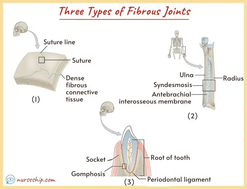
There are 3 types of fibrous joints. They are:
i. Sutures
Sutures are made up of relatively short connective tissue fibers. These fibers are continuous with the periosteum, forming hard structures that connect the skull bones together.
Sutures in the skull are found throughout the fetal and early childhood stages of development. It gradually closes as the child matures.
When the sutures are closed, it is referred to as syndesmoses.
ii. Syndesmoses
Ligaments join the bones in syndesmoses, and the connecting fibers are longer than those seen in sutures. The lengths of these fibers determine how much mobility is possible at these joints.
The ligament that joins the distal end of the fibula and tibia is an example of a syndesmoses joint. It is made up of extremely short fibers and have very little to no movement.
On the other hand, the syndesmoses joint, which connects the ulna and radius interosseous membranes (similar to ligaments), is longer and allows slightly higher joint flexibility.
iii. Gomphoses
The ‘gomphoses’ or ‘dentoalveolar’ joint is another type of fibrous joint found between teeth and jaws, which connects the teeth to their bony sockets.
The singular term gomphosis (plural: gomphoses) refers to how the teeth are embedded in their sockets.
Gomphoses are also called the peg-and-socket joint.
Function of fibrous joint
- They protect the internal organs
- They provide stability
Example of fibrous joint:
- Sutures in the skull
- Radioulnar joints
- Tibiofibular joints
2) Cartilaginous Joints
Cartilaginous joints are held together by hyaline cartilage or fibrocartilage. They are relatively firm but flexible connective tissue.
They are slightly movable joints with some degree of mobility and are also called Amphiarthrosis.
Cartilaginous joints are further classified as follows:
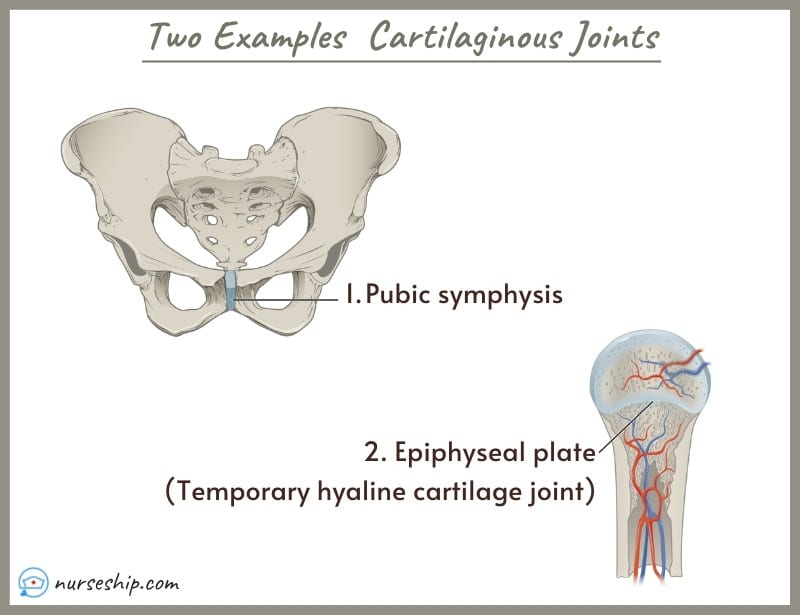
i. Synchondroses: The joints in which the cartilage is temporarily present in children during growth and is replaced by bone in adulthood.
For example, the epiphyseal plates in long bones.
ii. Symphyses (singular: symphysis): Permanent fibrocartilage is present in these joints.
An example of a symphysis joint is the pubis symphysis.
Function of cartilaginous joint
- They allow limited movements
- Serve as growth centers for long bones during childhood
Examples of cartilaginous joints:
- The spinal vertebrae are joined together by cartilaginous vertebral discs which provide limited mobility.
- The symphysis between pubic bones also allows little movement.
3) Synovial Joints
The synovial joints, also known as diarthrosis, are the most abundant and versatile type of joints present in the human body. They have the greatest range of flexibility and motion.
The movements in synovial joints are mainly angular movements that allow for a change in position by changing the angle between the bones.
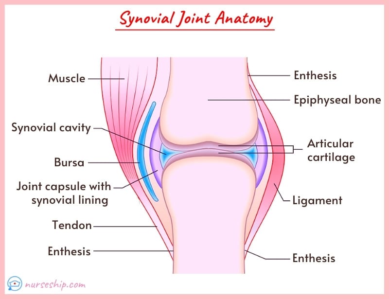
Distinct features of synovial joints include a joint cavity, synovial fluid, and the capsule.
Some synovial joints may have a shock-absorbing fibrocartilage pad called menisci (singular: meniscus) and fluid-filled sacs called bursae (singular: bursa). They are located in the space between tendons and the underlying bony prominences, such as the elbow or knee.
Bursae help to increase joint mobility and reduce friction.
What are the structures of the synovial joints?
Synovial joints are more complex than other types of joints and they have six distinct characteristics.
The following describes all six synovial joint structures.
1. Joint cavity:
The space between the articulating bones is known as a joint cavity.
2. Synovial fluid:
Synovial fluid is the colorless slippery liquid present in the joint cavity that lubricates the joint to make the movements easier.
3. Joint capsule:
The joint capsule is a sac-like capsule of connective tissue that encapsulates the joint cavity.
It extends from the periosteum of the articulating bones.
4. Synovial membrane:
The synovial membrane makes up the slippery inner layer of the joint capsule that produces synovial fluid.
5. Articular cartilage:
Articular cartilage is the hyaline cartilage lining the articular surfaces of bones in the synovial joints to allow smooth and frictionless movements.
6. Ligaments:
Ligaments are tough fibrous bands of connective tissue that bind the bones in joint capsules.
What are the types of synovial joints?
The synovial joints are responsible for a wide range of movements and they are not identical. They are classified based on their anatomy and movement patterns.
There are different 6 types of synovial joints. Namely,
- Gliding joint
- Hinge joint
- Pivot joint
- Condyloid joint
- Ball and socket joint
- Saddle joint
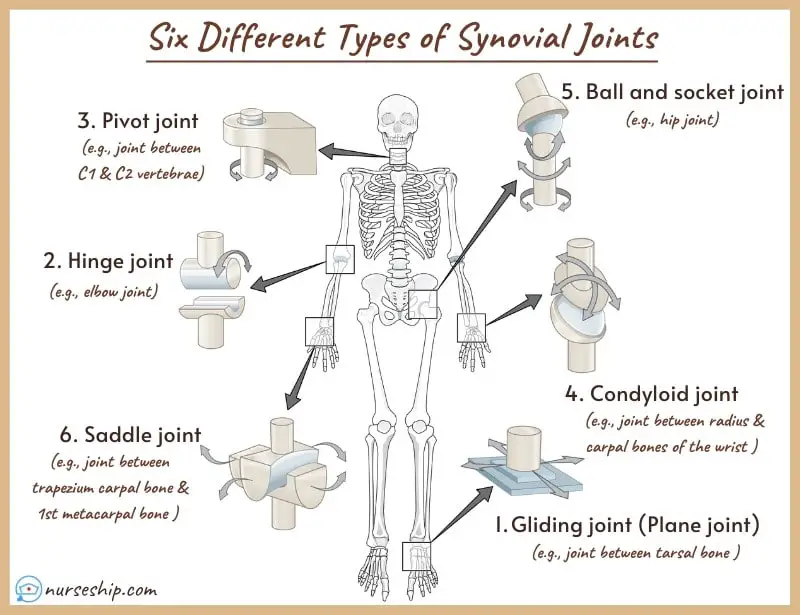
1. Gliding joint
Gliding synovial joints allow gliding movements due to the presence of flat or slightly curved articular surfaces.
The gliding movements are limited and directed by the ligaments. These are also called plane joints.
Example of gliding joints:
- Intercarpal joints of hands
- Intertarsal joints of feet
2. Hinge joint
Hinge joints work like the hinge of a door. In these joints, the convex surface of one bone fits with the concave surface of the other bone.
Back-and-forth movements are possible at these uniaxial hinge joints.
Example of hinge joints:
- Elbow joints
- Knee joints
3. Pivot joint
Pivot joints, as the name implies, rotate around a single axis. The projection of one bone articulates with the ring-shaped socket of the other bone.
Example of pivot joints:
- Joint between first and second cervical vertebrae
- Radioulnar joint
4. Condyloid joint
The condyloid or ellipsoid joints involve the articulation of the convex end of one bone with similar opposing depression on the other bone.
Flexion, extension, and side-to-side movements are possible at condyloid joints.
Example condyloid joints:
- Knuckles of hand
- Joint between the distal end of the radius and carpal bones of the wrist
5. Ball and socket joint
The ball and socket joint, as the name implies, involves the articulation of one bone’s ball-shaped head to the cup-like socket of the other bone.
The ball and socket joint allows the movement of distal bone in all planes and rotation around the central axis.
Example of ball and socket joint:
- Shoulder joint
- Hip joint
6. Saddle joint
In saddle joints, the bone surfaces have a concave form like a saddle. Saddle joints are similar to condylar joints.
Saddle joints can move around two axes thus allowing flexion, extension, adduction, and abduction. The side-to-side motion is limited as compared to the back-and-forth movement.
Example of saddle joint:
The only saddle joint present in the human body is the carpometacarpal joint i.e., the thumb joint.
Related Articles
20 different types of anatomical body movement in humans
Male Pelvis vs Female Pelvis Anatomy: What is the difference?
Summary
| The main function of the joints: To facilitate mobility |
| Three types of joints: 1. Fibrous joints (synarthrotic); 2. Cartilaginous joints (amphiarthrotic); and 3. Synovial joints (diarthrotic) |
| Three types of fibrous joints (synarthrotic): 1. Sutures; 2. Syndesmoses; and 3. Gomphoses |
| Two types of cartilaginous joints (amphiarthrotic): 1. Synchondroses; and 2. Symphyses |
| Six types of synovial joints: 1. Gliding joint; 2. Hinge joint; 3. Pivot joint; 4. Condyloid joint; 5. Ball and socket joint; and 6. Saddle joint. |
| Six characteristics of synovial joints: 1. Joint cavity; 2. Synovial fluid; 3. Joint capsule; 4. Synovial membrane; 5. Articular cartilage; and 6. Ligaments. |
Conclusion
To conclude, the main function of the human body’s joints or articulations is to facilitate mobility. There are different types of joints, which are classified based on their function and accompanying structures.
References
Lippincott Williams & Wilkins. (2009). Anatomy and physiology made incredibly easy! (3rd ed.).
Cohen, B., & Taylor, J. (2009). Memmler’s Structure and Function of the Human Body, 8th ed (10th ed.). Lippincott Williams & Wilkins.
Moini, J. (2020). Anatomy and Physiology for Health Professionals (3rd ed.). Jones & Bartlett Learning, LLC. Thompson, G. (2015). Understanding Anatomy & Physiology: A visual, auditory, interactive approach (2nd ed.). F. A. Davis Company.