
Inversion vs Eversion in Anatomy
The eversion and inversion are complex movements involving the many plane (gliding) joints between the tarsal bones of the posterior foot (intertarsal joints). Sometimes, they also occur at the ankle joint.
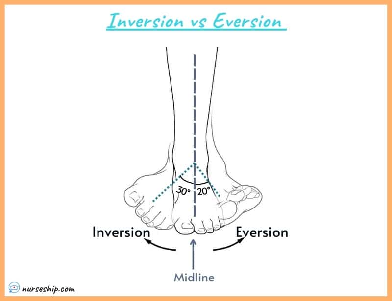
Eversion and inversion movements occur in relation to the midline of the body.
The movement of the foot that turns the soles inwards is called inversion, while the opposing movement is called eversion.
The foot can move more in an inverted position than in an everted position.
Eversion and inversion movements support the foot’s stability when walking or running on an uneven surface. Also, these movements facilitate fast side-to-side direction changes in athletic activities like racquetball, basketball, or soccer.
Continue reading to learn more about inversion vs eversion; muscles, and nerves responsible for these movements.
What is an inversion in anatomy?
In anatomy, inversion is the movement that causes the sole of the foot to rotate in a medial direction, towards the midline of the body.
The fact that the term “in” comes before “inversion” makes it obvious that the sole is pointed inward (medially).
The normal range of motion for inversion of the foot at the ankle is 0 to 30 degrees.
Example of inversion:
When you twist your ankle, you found your foot rotated in an inward direction.

Human Anatomy & Physiology Course
- Guidebooks
- Complete Diagrams & Lessons
- Certifications
- Quiz & Answers
- And More…
What is eversion in anatomy?
In anatomy, foot eversion is defined as the movement in which the foot’s sole rotates away from the midline of the body creating an opposing motion (laterally).
Eversion is a literal meaning of the word “evert,” which means to “turn outward.”
The normal range of motion for eversion of the foot at the ankle is 0 to 20 degrees.
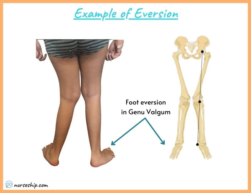
For example:
When a person walks with Genu Valgum, you can see slight eversion on his/ her foot.
Inversion and eversion of the foot
Muscles and nerve supply
The muscles responsible for the inversion of the foot are the tibialis posterior and tibialis anterior.
Both the deep peroneal nerve and the tibial nerve supply the tibias anterior and tibialis posterior muscles, respectively.
On the other hand, eversion of the foot is caused by the muscles fibularis longus, fibularis brevis, and fibularis tertius.
The superficial fibular nerve supplies these evertors muscles.
The movements of eversion and inversion of the foot occur primarily at the Talocalcaneonavicular joint and the Subtalar joint (aka, Talocalcaneal joint).
Inversion and eversion of the ankle
Muscles and nerve supply
Inversion of the ankle is performed by tibialis anterior, flexor digitorum, tibialis posterior, flexor hallucis, and extensor hallucis.
The nerve supply of these muscles is as follows, Tibialis anterior is supplied by the deep peroneal nerve, and the Tibialis posterior is supplied by the tibial nerve.
Similarly, flexor hallucis by the tibial nerve, extensor hallucis by the deep peroneal nerve, and nerve supply of flexor digitorum is the tibial nerve.
The peroneus brevis and peroneus longus facilitate the eversion of the ankle, and superficial fibular nerve or superficial peroneal nerve supplies these muscles.
The other muscles such as extensor peroneus tertius and digitorum longus also contribute to ankle eversion and are innervated by the deep peroneal nerve.
The ankle inversion and eversion occur at the subtalar joint.
Calcaneal inversion vs eversion
The major difference between calcaneal inversion and calcaneal eversion is that during an open kinetic chain inversion, the calcaneum rolls into an inverted position and moves to the side laterally.
While during eversion, the calcaneum rolls into eversion and slides toward the middle of the foot.
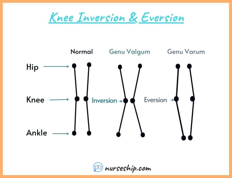
Knee eversion vs inversion
Normally, the movements of eversion and inversion do not occur at the knee joint. But there are two conditions in which the knee is said to be inverted or everted in position. These two conditions are genu valgum and genu varum.
Genu valgum is a condition in which a person stands up straight with their knees touching but their ankles apart. This condition is also called genu valgum.
On the other hand, genu varum also known as bowed legs is a condition in which the knee bends outward while the feet and ankle touch together.
Inversion and eversion: Ankle sprains
An eversion sprain occurs when the ankle rolls in an outward direction and ruptures the deltoid ligaments (medial ligament), this is called an eversion ankle sprain.
On the other hand, inversion ankle sprains occur when the sole of the foot turns too far inward, hurting the ligaments on the foot’s lateral side.
Inversion sprains are more common but are less severe than eversion sprains.
The most commonly injured ligaments in the case of inversion sprain are” anterior talofibular” (ATFL), the “calcaneofibular” (CFL), and “posterior talofibular” (PTFL) ligaments.

How do you remember inversion and eversion?
The 2nd letter in the word eVersion is a “V“, which makes it easy to remember. When you turn both feet outward, the outside edges of both feet will lift off the ground, making a “V” shape with your feet.
On the other hand, the tip to remembering INversion is to focus on the word “IN”, in means inside, inward, inside is the medial side and eversion will be opposite.
Here is a tip to remember the muscles responsible for inversion and eversion as well.
Note that the initial letter of the movement’s name is the same as the second letter of the muscle’s name.
Inversion
- 1st letter of the movement is “I”: Inversion (Invertor muscles [muscle group name])
- 2nd letter of the muscle is “I” = tIbialis anterior and tIbialis posterior.
Eversion
- 1st letter of the movement is “E”: Eversion (Evertor muscles [muscle group name])
- 2nd letter of the muscle is “E” = pErineus Tertius, pErineus Brevis, and pErineus longus.
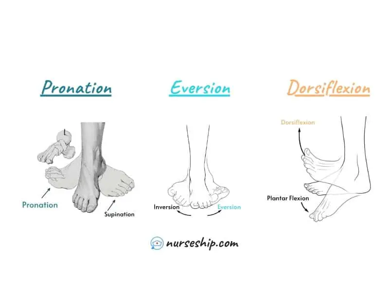
Is pronation inversion or eversion?
Sometimes, eversion is called pronation. The only difference between these two movements is that eversion occurs in the frontal plane while pronation is a tri-planar movement that occurs in three planes.
Pronation is made up of abduction of the forefoot (frontal plane), dorsiflexion of the talocrural (ankle) regions (sagittal plane), and eversion of the hindfoot (transverse plane).
Related Articles
20 Different Types of Anatomical Body Movements |Examples |Illustrations
Different types of joints in the human body
Male Pelvis vs Female Pelvis Anatomy: What is the difference?
Inversion and eversion in anatomy: A summary
| Eversion | Inversion |
| Sole of the foot is turned outward to face laterally | Sole of the foot is turned inward to face medially |
| Movement occurs at the Talocalcaneonavicular and Subtalar joint | Same joints (i.e., Talocalcaneonavicular and subtalar joint) |
| Muscles responsible for eversion movement are fibularis brevis and longus | Muscles responsible for inversion movement are tibialis posterior and tibialis anterior |
| Normal range of motion for eversion of the foot at the ankle is 0 to 20 degrees | Normal range of motion for inversion of the foot at the ankle is 0 to 30 degrees |
Conclusion
To conclude, eversion is the movement in which the foot rolls in an outward direction but in case of excessive eversion motion like in eversion ankle sprain ligaments on the opposite side, that is the medial ligaments are damaged.
The opposite movement of eversion is inversion.
Reference
Moini, J. (2020). Anatomy and Physiology for Health Professionals (3rd ed.). Jones & Bartlett Learning, LLC.
Moore, K., Dalley, A., & Agur, A. (2018). Clinically Oriented Anatomy (8th ed.). 8 Wolters Kluwer.
Thompson, G. (2015). Understanding Anatomy & Physiology: A visual, auditory, interactive approach (2nd ed.). F. A. Davis Company.
Wineski, L., & Snell, R. (2019). Snell’s Clinical Anatomy by Regions (10th ed.). Wolters Kluwer.